



Image-Guided Surgery
Real-time coronary stenosis detection using deep learning for intraoperative angiographic image analysis with 0.94 mAP.
April 2021
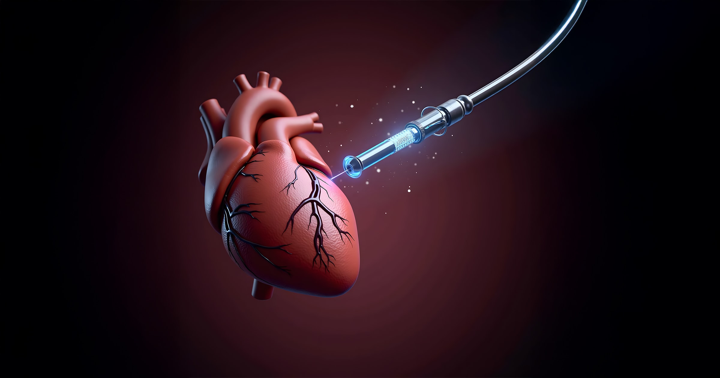
Real-time landmark tracking for safer valve implantation during TAVI procedures
The Kemerovo Cardiology Center needed better intraoperative guidance for TAVI procedures to reduce valve misplacement risks.
Develop an AI-driven visual assistance system for real-time surgical support during transcatheter aortic valve implantation.
Applied multi-task deep learning to detect and localize 11 anatomical keypoints from aortography data using ResNet and MobileNet V2.
Delivered 97% accuracy and ~90 FPS speed, improving precision and enabling safer, future-ready valve implantation.
This project addresses one of the most critical challenges in modern cardiovascular intervention: accurately positioning the valve during Transcatheter Aortic Valve Implantation (TAVI). Traditional imaging methods offer limited intraoperative guidance, risking misplacement and complications.
To overcome this, we developed a deep learning-based visual assistance system using multi-task learning (MTL) to simultaneously detect and localize 11 anatomical keypoints on both the aortic valve and the delivery system. This system was trained on real intraoperative aortography data and evaluated across several neural network architectures. The most efficient models (ResNet and MobileNet V2) achieved impressive accuracy and responsiveness, demonstrating real-time prediction capabilities that could enhance clinical outcomes and serve as the basis for future robotic surgical systems.
The solution was developed and validated using real-world surgical imaging data, making its outputs directly relevant for clinical applications. The dataset included:
These images captured three critical surgical stages providing a diverse and representative training ground for keypoint detection (Figure 1).
The project implemented a multi-task learning architecture to simultaneously classify the presence and regress the coordinates of keypoints in angiographic images (Figure 2). This approach offered more efficient representation learning compared to single-task methods.
The system achieved real-time performance with high accuracy in both keypoint detection and localization. Key outcomes included:
The analysis also highlighted challenges from class imbalance, particularly with underrepresented keypoints (e.g., aortic annulus landmarks), which the model handled well despite data limitations. Training convergence for selected architectures is shown in Figure 3, and prediction quality comparison in Figure 4.
MobileNet V2
ResNet V2
Inception V3
This project successfully demonstrates a real-time AI-based system for intraoperative landmark tracking during TAVI, offering a significant step toward safer, more precise cardiovascular surgery. The system's main contributions are:
Future directions include expanding the dataset to cover more valve models, optimizing for class imbalance, and integrating with surgical robotics for semi-automated procedures.
Real-time coronary stenosis detection using deep learning for intraoperative angiographic image analysis with 0.94 mAP.
AI-powered microscopy pipeline for detecting and counting cancer cell biomarkers using EfficientDet, achieving 85% mAP for nuclei detection.