



Biomedical Research
AI-powered microscopy pipeline for detecting and counting cancer cell biomarkers using EfficientDet, achieving 85% mAP for nuclei detection.
January 2021
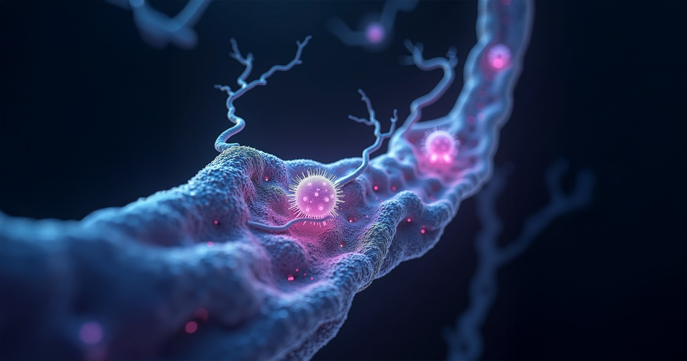
Microvascular feature segmentation in tissue-engineered vascular grafts
Clinical biologists at the Kemerovo Cardiology Center needed accurate segmentation of microvascular features in TEVG histology images to assess vascular regeneration in sheep models.
Lead model development and deliver a robust segmentation pipeline for nine histological features.
Optimized six deep learning architectures and created an ensemble model using cross-validation, augmentation, and Bayesian tuning.
Achieved a mean Dice score of 89%, enabling precise tissue analysis and accelerating biomedical research.
This project focused on developing a deep learning-powered pipeline for segmenting and quantifying histological features in tissue-engineered vascular grafts (TEVGs). Using advanced deep learning models, the study aimed to address challenges in analyzing complex tissue regeneration patterns within biodegradable grafts implanted in sheep carotid arteries.
By optimizing six neural networks and creating an ensemble model, the project achieved precise segmentation of key microvascular features, including arterioles, venules, capillaries, immune cells, and nerve trunks. The ensemble model demonstrated state-of-the-art accuracy, achieving a mean Dice Similarity Coefficient (DSC) of 0.889, offering a robust tool for accelerating research in tissue engineering and translational medicine.
The study utilized a dataset comprising 104 Whole Slide Images (WSIs) obtained from biodegradable TEVGs implanted into the carotid arteries of 20 sheep. After six months, the sheep were euthanized to assess vascular tissue regeneration patterns.
The methodology was designed to address the complexity of segmenting microvascular features in tissue-engineered vascular grafts by combining advanced deep learning techniques with rigorous evaluation protocols:
The results demonstrate the effectiveness of the proposed pipeline in accurately segmenting microvascular features of TEVGs. Individual models performed well across various features, while the ensemble model significantly improved overall accuracy:
The ensemble of three models (MA-Net, DeepLabV3, and FPN) provided the optimal solution (Figure 3).
This project demonstrated the effectiveness of deep learning models in segmenting microvascular structures within tissue-engineered vascular grafts. By integrating advanced algorithms like MA-Net and DeepLabV3, the workflow achieved high precision in analyzing tissue regeneration patterns.
These findings pave the way for enhanced automation in tissue engineering research. Future work could explore extending this pipeline to analyze more complex vascular networks or incorporating foundation models for broader applicability.
AI-powered microscopy pipeline for detecting and counting cancer cell biomarkers using EfficientDet, achieving 85% mAP for nuclei detection.
Explainable AI framework for detecting pulmonary edema features in chest X-rays using deep learning segmentation and object detection.